


Click here to view a Non-Invasive Vascular Ultrasound
What is circulation (vascular) ultrasound?
 Circulation (Vascular) Ultrasound provides your doctor with moving images of your circulatory system (arteries and veins), and takes excellent pictures that will help your doctor to evaluate your circulation’s health. A specially trained technician (Sonographer) will use a gel to slide a microphone-like device called a transducer over your body. Reflected sound waves will provide images of your veins and arteries. Circulation (Vascular) Ultrasound uses the same technology that allows doctors to see an unborn baby inside the pregnant mother. In some cases, a special dye may need to be injected into the vein to help enhance the images so that your doctor can better evaluate the health of your circulatory system. This ultrasound does not involve radiation.
What is Peripheral Vascular Ultrasound?
This procedure uses sound waves to obtain images and measure speed (velocity) of blood flow in carotids (neck), arms, legs, abdominal aorta, and renal (kidney) blood vessels. These images are analyzed to determine whether or not you have blockages in your arteries, blood clots in your veins, or if an abdominal aortic aneurysm is present.
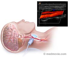
What is Carotid Ultrasound?
This procedure uses sound waves to obtain color images of the arteries in your neck. The physician evaluates the images to determine to what extent these arteries are blocked and how much blood is flowing to your brain and eyes. There are two carotid arteries, one on each side of your neck. Both sides will be checked during the procedure. This test takes one hour and no preparation is needed.
Last Update 5/27/2011


