


What are the types of Heart Ultrasound?
What types of echocardiograms are there?
The most common type of echo performed is the transthoracic echo, which is performed by placing the probe on the outside of the chest wall with a gel-like substance to transmit sound waves into the body. There are also several other types:
Doppler Echocardiogram
Evaluate blood flow in the heart and blood vessels. This procedure measures the speed and direction of the blood flow within the heart. It screens the four valves for leaks. With Doppler echocardiograms, as the wand moves over your heart you will hear a “whooshing” sound much like that of a washing machine. This is the sound of blood moving within your heart.
The stress echo or stress test
Combines the echo exam with a treadmill to walk on or bike to pedal, or medication that shows the effect of exercise on the heart. Stress tests are used to diagnose the narrowing of the coronary arteries.
The Contrast Echocardiogram
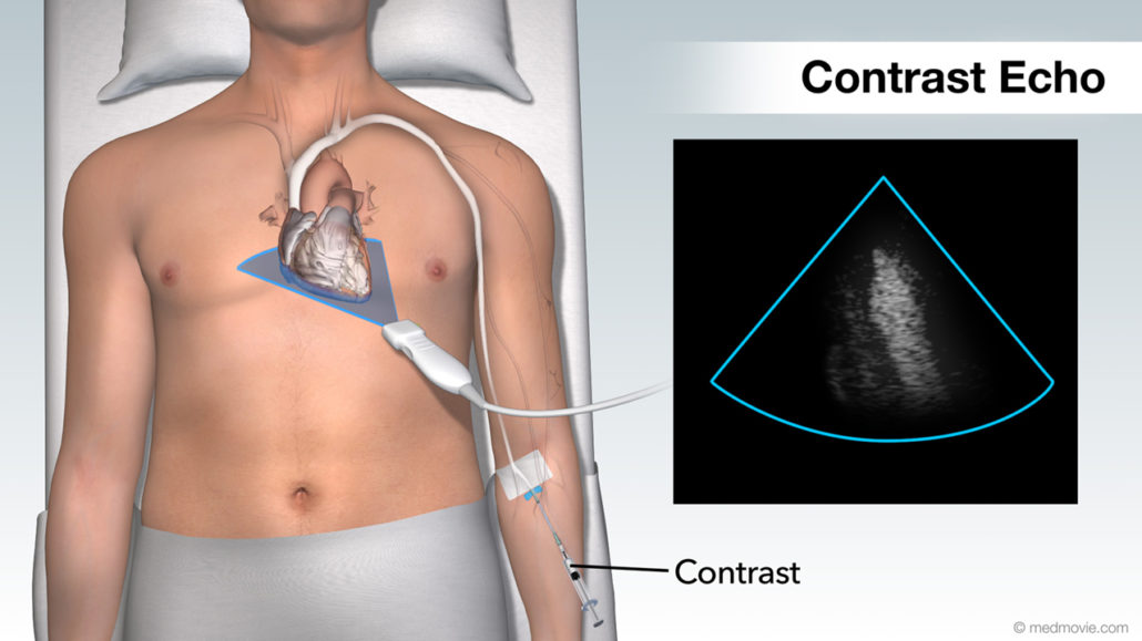
Click the image to view an animation of Contrast Echocardiogram
Combines an echocardiogram with an IV that contains a solution which allows the sonographer or physician to see the inside of the heart more clearly. This solution is injected into your vein through an IV. Your doctor will discuss with you whether contrast is needed, and what, if any, possible side effects the contrast solution could have. Thousands of patients have successfully experienced a contrast echocardiogram, however, in rare cases side effects can occur in certain individuals.
Transesophageal echo
is a form of echo where a miniature ultrasound camera is passed down the throat to locate the back of the heart. This allows the physician to obtain very high-quality moving images. You can be sedated during the procedure if you wish. Transesophageal echocardiograms are typically performed to evaluate serious heart conditions.
3D Heart Ultrasound
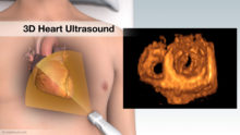
Click the image to view an animation of 3D Heart Ultrasound
Provides your doctor with moving images of your heart and takes excellent pictures that will help your doctor evaluate your heart health. A specially trained technician, (Cardiac Sonographer), uses a gel to slide a microphone-like device called a transducer over the chest area. This allows reflected sound waves to provide a live 3D picture of your heart and valves. 3D Heart Ultrasound uses the same technology that allows doctors to see an unborn baby inside a pregnant mother. No radiation is involved in heart ultrasound, and the technology can be used on people of all ages.
The Most Common Heart Ultrasound: Transthoracic Echocardiogram
FREQUENTLY ASKED QUESTIONS ABOUT HEART ULTRASOUND (ECHOCARDIOGRAPHY)
Click here to view a Heart Ultrasound Echocardiogram
Q: What is a Heart Ultrasound (Transthoracic Echocardiogram; TTE; Echocardiogram; Echo)?
A: A heart ultrasound is a useful tool to evaluate the structure and function of the heart and associated vessels. It is a fast, easy and painless evaluation that uses ultrasound waves to produce images of the heart. In North America, the test is performed by a specially trained technologist, called a sonographer, and is interpreted by a specially-trained physician, usually a cardiologist, trained in reading heart ultrasounds. Heart Ultrasounds provide your doctor with moving pictures of your heart that allows your doctor to evaluate your heart’s health. This ultrasound uses the same technology that allows doctors to see an unborn baby inside a pregnant mother.Q: Why has my doctor requested that I have a heart ultrasound (echocardiogram)?
A: There are many reasons that your physician may request that you have a heart ultrasound. Physicians use it evaluate your heart’s performance as well as the structures of the heart, including the heart chambers and valves. An echo may sometimes also be used to look for the cause of a murmur, to check the size of the heart chambers, to check for fluid around the heart, or to inspect the pumping capability (the muscles) of the heart if a patient has short of breath or has complained of certain symptoms during exertion.Q: How do I prepare for an echo?
A: There is no special preparation required for a heart ultrasound. You should come as you are and eat or drink as you normally do. If you take medications, you should continue to take them as normal unless your doctor specifies otherwise. You should plan on being at the Echocardiography Lab for about forty-five minutes to one hour.Q: What should I expect?
A: Upon arrival at the lab the friendly staff will greet you. They may request you to provide additional insurance information, and will ask you to register. They may also ask that you provide a prescription or order for your exam.You will then be escorted into an ultrasound room. The room will be dimly lit and will contain an examination table or bed and an ultrasound machine. You may be asked a few questions by the sonographer who will want to know why you are having the test, if you have had previous tests, and if you have ever had open heart surgery. Usually a brief explanation of the procedure will be given as well.
You will be asked to remove your clothing from the waist up and women will be given a gown to wear during the procedure
If you need help, the sonographer will assist you in getting onto the exam table, where you will be asked to lie on your left side. The sonographer will then attach ECG lead wires to electrodes adhered to your chest with simple medical tape.
The lights may be dimmed to allow the sonographer to see the monitor better.
The sonographer will apply ultrasound gel to a microphone-like device called a transducer. The transducer sends and receives the harmless ultrasound waves. The gel allows the ultrasound beams to penetrate your chest wall so that it is possible to “see” your heart.
Next, the sonographer will begin to acquire ultrasound images and audio recordings by methodically and precisely moving the transducer around on your chest, stomach, and neck. The sonographer will be viewing these images on a monitor and will take various recordings at several different locations or “views”. During recording, you may be asked to change your position and to hold your breath. These variations in position and breathing allow the Sonographer to obtain the best quality pictures. The Sonographer will press the transducer against your skin and this pressure may be moderate at times to facilitate the transmission of ultrasound waves. If it becomes uncomfortable, please let the sonographer know.
You should try to remain still and quiet during the exam. The imaging will take about 30 to 45 minutes. Often, the Sonographer will review the study with a supervisor or physician while you are still in the room. You should not be alarmed; the technique to acquire images of the heart is technically demanding and sonographers frequently rely on the advice of others as they acquire these images.
The images and sounds of the exam will be recorded on a computer disk and/or videotape for later review.
Q: What will I see and hear on the heart ultrasound machine during my exam?
A: Ultrasound waves used in performing the echocardiogram are not audible to the human ear, so you will not hear the sound waves.
Structures will be displayed in “real-time” and appear as white moving objects on the screen. For example, the valves of the heart will look like white flap-like moving structures. Areas of the heart where there is fluid or blood look black on the screen.During the exam, you will notice the sonographer placing marks on the screen with small computer calipers. The sonographer uses the calipers to perform various measurements of the size, function and blood flow of the heart.
An echocardiogram exam usually includes a Doppler recording of the blood movement or flow within the heart. When color flow Doppler is used in the exam it will appear as different colors moving within the white and black images on the monitor. The different colors represent the different speeds and directions of blood flow in the heart.
Doppler examinations often also include an audio signal of the blood flow. These audio signals can be heard and seen. During the audio Doppler recording, you will hear the sound of the blood moving through the heart and the sound of the heart valves opening and closing. The audio signals are also displayed as a graph on the monitor. These graphic recordings help the physician to determine valve function and heart pressures.
Q: What happens after the exam?
A: Following the recording of the images, the Sonographer will remove the ECG electrodes from your chest, will wipe off the ultrasound gel from your skin, will help you off the examination table and escort you out of the lab.The ultrasound images and Doppler recordings will be submitted to a Cardiologist who is a specially-trained physician in reading heart ultrasounds. He or she will interpret the images and will then provide your general physician with a written report. Often, you will not be given any results for one or two days. Generally, the Sonographer will not provide you with any results at the time of the examination.
Frequently Asked Questions about Exercise Heart Ultrasound (Stress Echocardiogram)
Q: What is an Exercise Heart Ultrasound (Stress Echo)?
A: An Exercise Heart Ultrasound, (Stress Echocardiogram), sometimes called a stress echo, is a tool used to evaluate heart function by combining an exercise (stress) test with a heart ultrasound. Exercise Heart Ultrasound uses ultrasound waves to produce images of the heart both before (sometimes during) and immediately following exercise. Images of the heart at rest are compared with images of the heart during and/or after exercise to evaluate how the heart responds to exercise.
In North America the test is performed by a specially trained technologist, called a sonographer, and is interpreted by a physician trained in reading exercise heart ultrasounds. During the exercise portion of the exam there will be a medical staff member, usually a physician, supervising the examination and there is sometimes a third person assisting.Patients that have physical limitations that cause them to be unable to exercise may be given a pharmacologic stress heart ultrasound (where a medication that simulates the effects of exercise is given) instead of an exercise heart ultrasound.
Q: Why has my doctor requested that I have an exercise heart ultrasound?
A: If you are going to have an exercise heart ultrasound, it is more than likely that it has been requested or ordered by your physician or cardiologist. This type of ultrasound is most often requested to check for good blood flow to the heart. It may help to provide an early detection of coronary artery blockage.Q: What must I do to prepare for an exercise heart ultrasound?
A: Your doctor and/or the laboratory where you will have the ultrasound performed will provide you with written instructions to help you to prepare for your appointment. You may be asked to refrain from eating and drinking for a few hours before the exercise heart ultrasound and you may also be asked to limit your drinking to water and other caffeine free drinks for up to 24 hours before your appointment You may be asked to temporarily hold off on taking some medications. You may also be asked to stop using tobacco products for a few hours beforehand.You should bring or wear comfortable walking shoes and wear suitable (warm-ups/loose fitting clothes). It is very important that you check with your doctor and/or the heart ultrasound laboratory staff a few days before your appointment for any specific instructions. You should plan on being at the heart ultrasound (echocardiography) lab for anywhere from one to two hours.
Q: What should I expect while at the exam?
A: Upon arrival at the lab you will be greeted by lab staff. They may need to obtain some insurance information from you. You will be asked to register and may also be requested to provide a prescription or order for your examination.After processing these items, you will be escorted into an examination room. The room will have a special examination table, a heart ultrasound machine and a treadmill (or sometimes a stationary bicycle).
You may be asked a few questions by the sonographer who will want to know why you are having the test, if you have had any previous exercise heart ultrasounds, and if you have ever had open heart surgery. Usually he/she will give you an explanation of the procedure also.
You will then be asked to remove your clothing from the waist up and will be given a gown to wear.
A staff member will attach ECG lead wires to electrodes attached to your chest with simple medical tape. To ensure the ECG lead wires stay attached while you exercise, we will clean your skin and prepare it for the electrodes. For men, it may be necessary to shave small areas of chest hair.
A blood pressure cuff will be placed on your arm before you begin to exercise.
You may be asked to have an intravenous line (IV) started so that a special echocardiographic contrast material can be used to enhance the ultrasound pictures. The lab performing the exam will explain this procedure if they feel it will be beneficial.
The sonographer will obtain resting images before you begin to exercise.
The lights will be dimmed to allow the sonographer to see the monitor better.
You will be asked to lie on the bed on your left side.
The sonographer will apply ultrasound gel to a microphone-like device called a transducer. The transducer sends and receives the harmless and painless ultrasound waves. The gel allows the ultrasound beams to penetrate your chest wall to your heart and makes it possible to “see” the heart.
The sonographer will then begin to acquire ultrasound images by methodically and precisely moving the transducer around on your chest and abdomen. The sonographer will be viewing these images on a monitor and will take various recordings at several different locations or “views”. During the recording you may be asked to change your position and to hold your breath. These variations in position and breathing allow the sonographer to obtain the best quality pictures possible. The sonographer will press the transducer against your skin and this pressure may be moderate at times to facilitate the transmission of ultrasound waves. If it becomes too uncomfortable, please let the sonographer know and he/she will let you take a short break. After the sonographer has obtained all of the resting images, the exercise portion of the exam will begin.
ECG and blood pressure readings will be taken before exercise begins and throughout the exercise portion of the test.
Usually exercise is performed on a treadmill, or less commonly, on a stationary bicycle. When using the treadmill, the treadmill will begin at a slow, warm-up speed. The speed and the incline (or slope) of the treadmill will be increased every few minutes throughout the exercise heart ultrasound.The exercise heart ultrasound is most useful if your target heart rate is reached before exercise is stopped so your doctor will want you to exercise as long as you can. Exercise will usually continue until your target heart rate is reached unless you experience difficulty breathing, or other show other symptoms, and they will immediately stop.
IT IS IMPORTANT THAT YOU IMMEDIATELY LET YOUR DOCTOR KNOW IF YOU EXPERIENCE ANY CHEST PAIN, SEVERE SHORTNESS OF BREATH, DIZZINESS, OR OTHER SYMPTOMS DURING THE EXAM.
More images of your heart will be obtained immediately after exercise is stopped (if you exercise on a stationary bicycle images may also be obtained during exercise).
After the treadmill is stopped, you will be escorted very quickly back onto the bed, and asked to lie on your left side. It is important that you are positioned on the bed very quickly so that we can capture images of your heart while your heart rate is still high.
The sonographer will obtain another set of images so that the resting images before you began exercise can be compared to the images of your heart when it is under stress (immediately after you have exercised).You will be asked to remain on the bed until your heart rate has slowed. If you experience any symptoms during this time or at any other time, you should report them immediately to the staff.
The resting and exercise images of the exam will be recorded on a video tape and/or computer disk for later review and analysis by a cardiologist/echocardiographer, who is a physician in specially-trained reading and interpreting ultrasound images.
Q: What happens after the exam?
A: Following the recording of the images, the sonographer will remove the blood pressure monitoring cuff from your arm and the ECG electrodes from your chest, will wipe off the ultrasound gel, will help you off of the bed and will escort you out of the lab.
The ultrasound images will be interpreted by the Cardiologist (this may be the physician that was present during the stress echo). He or she will interpret the stress echocardiogram and will provide your general physician with a written report. Sometimes the physician present during your stress echocardiogram will be able to tell you his or her preliminary findings although this immediate feedback varies depending on individual circumstances.
Frequently Asked Questions about Transesophageal Echocardiograms
Q: What is a Transesophageal Echocardiogram (TEE)?
A: A Transesophageal Echocardiogram is a useful tool used to evaluate the function and small detailed structures of the heart and associated vessels. The Transesophageal Echocardiogram is a variation of the Transthoracic Echo procedure. The TEE procedure uses ultrasound waves to produce images of the heart. Performing a TEE involves passing a tube into the esophagus, or swallowing tube. In North America the test is performed and interpreted by a physician, usually a cardiologist, specially trained in Transesophageal Echocardiography
Q: Why has my doctor requested that I have a Transesophageal Echocardiogram?
A: If you are going to have a TEE it is more than likely that it has been requested or ordered by your physician or a cardiologist. There are many reasons for requesting that you undergo this procedure. Physicians use this procedure to look for any abnormalities in the physical structures of the heart, including the heart chambers, valves, and associated blood vessels. This procedure is sometimes also used to look for abnormalities within the structures that a standard transthoracic echocardiogram is not able to highlight. This may include examining the detailed structures and functioning of the heart valves.Q: How do I prepare for a transesophageal echocardiogram?
A: A transesophageal echocardiogram involves passing a narrow tube into the esophagus or swallowing tube. This requires a number of special preparations you may need to complete before the procedure. These preparations vary depending on the lab performing the procedure, so you should check with the lab performing your transesophageal echocardiogram. In general, you should not eat or drink fluids for several hours before the test, have someone drive you home, and be prepared to spend a few hours at the lab. If you currently take medications, you should check with your physician and/or the lab performing the test to let you know if you can continue to take them before the procedure.Q: What should I expect?
A: Upon arrival at the lab, our staff will greet you. We may need to obtain additional insurance information and you will be asked to register, and may need to provide a prescription or order for your examination.You will then be escorted into an ultrasound examination room. The room will be dimly lit and will contain a special examination table, an ultrasound machine, and other monitoring devices. You may be asked a few questions by the nurse or sonographer who will want to know why you are having the test, if you have had previous tests, if you have ever had open heart surgery and other questions about your medical history. You may be given a brief explanation of the procedure also. Sometimes, labs will call, or meet with you before the exam to ask these questions and explain the procedure.
You will then be asked to remove some of your clothing from the waist up. Women will be given a gown. If you need help, the sonographer or nurse will assist you in getting onto the stretcher. The staff will attach ECG lead wires to electrodes to your chest. These are attached using basic medical tape. A blood pressure cuff will be placed on your arm and a simple finger clip oxygen sensor will also be attached. You will be given oxygen through a tube which is worn on your nose. We will start an intravenous line (IV). If you wear dentures or partials, we will ask you to remove them.
The physician or nurse may give you IV medications to help you relax during the procedure. A topical medicine in the form of a gel or spray will be used to numb the back of your mouth.
The physician will place the flexible tube in your mouth. You will then be asked to swallow which will draw the tube into your esophagus. The tube is a modified endoscope scope which contains an ultrasound transducer that sends and receives the harmless ultrasound waves.
The physician will then begin to acquire ultrasound images and audio recordings by methodically and precisely moving the transducer in your esophagus and stomach. The movement of the tube should not be uncomfortable. The physician will be viewing the images on a monitor and will take various recordings at several different locations or “views”. You should try to remain still and quiet during the exam. The imaging will take 10 – 30 minutes.
The images and sounds of the exam will be recorded on a computer disk and/or videotape for later review.
Q. What will you see and hear on the echocardiography machine during your exam?
A. You may be able to see some of the images on the video monitor during the procedure.
Ultrasound waves used in performing the echocardiogram are not audible to the human ear, so you will not hear the sound waves. Structures will be displayed in “real-time” and appear as white moving objects on the screen. For example, the valves of the heart will look like white flap-like moving structures. Areas of the heart where there is fluid or blood will appear black on the screen.During the exam, you will notice the physician placing marks on the screen with small computer calipers. The physician uses the calipers to perform various measurements of the size, function and blood flow of the heart.
An echocardiogram exam usually includes a Doppler recording of the blood movement or flow within the heart. When color flow Doppler is used in the exam, it will appear as different colors moving within the white and black images on the monitor. The different colors represent the different speeds and directions of blood flow in the heart.
Doppler examinations often include an audio signal of the blood flow. These audio signals can be heard and seen. During the audio Doppler recording, you will hear the sound of the blood moving through the heart and the sound of the heart valves opening and closing. (This is the typical blub sound you have probably heard when listening to another person’s heart or on TV). The audio signals are displayed as a graph on the monitor. These graphic recordings help the physician to determine valve function and heart pressures.
Last Update 5/27/2011


